Empyema Thoracis
What is empyema?
Empyema is a collection of pus in the cavity between the lung and the membrane that surrounds it (pleural space).
It is caused by an infection that spreads from the lung and leads to an accumulation of pus in the pleural space. This infected fluid can build up which puts pressure on the lungs causing it to collapse leading to symptoms of shortness of breath and pain. The commonest cause of empyema in our country is Tuberculosis. It however can also be a complication of pneumonias, trauma with collection of clooted blood inside the chest that gets infected. Repeated aspiration of pleural fluids in cases of recurrent pleural effusion can lead to infection of the fluid that can lead to empyema. Finally empyema can happen after lung or chest surgery also.
Empyema Thoracis
What are the symptoms and diagnosis of empyema?
Symptoms
• Dry cough or Cough with sputum
• Fever and chills
• Excessive sweating, especially night sweats
• General discomfort, uneasiness or ill feeling (malaise)
• Unintentional weight loss
• Chest pain, which worsens on deep inhalation (inspiration)
Diagnosis
The diagnosis of empyema thoracis is suspected based on a detailed evaluation by a doctor. It can be confirmed using the below mentioned tests:
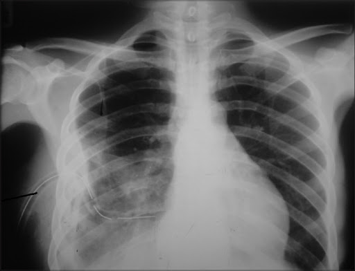
• Chest X-ray
• Thoracentesis
• Pleural fluid Gram stain and culture, Microbiological tests for TB
• CT scan of chest
Empyema Thoracis
What are the stages and treatments of empyema?
The aim of the treatment in cases of empyema is remove the infected fluid, cure the infection with appropriate antimicrobial therapy and ensure complete expansion of the lungs with no residual space or fluid.
Therefore, the treatment of empyema depends on the stage at which it presents to the doctor.
Empyema can progress through three stages if a person does not receive treatment.
Stage 1: Simple (the exudative phase)
The first stage of empyema is called simple empyema. It occurs when extra fluid begins to build up in the pleural cavity. This fluid can become infected and may contain pus. At this stage an effective drainage of the fluid either by thoracocentesis or chest tube drainage along with antibiotics may cure the disease.
Stage 2: Complicated (the fibrinopurulent phase)
In complicated empyema, the fluid in the pleural cavity begins to thicken and form “pockets.” At this stage an effective clearance of the infected fluid can be easily accomplished using thoracoscopic / VATS drainage of empyema. In this technique the surgeon makes three small holes in the chest through which he inserts a specialised telescope with attached camera and views inside of the chest cavity. With two additional instruments, the surgeon then breaks all the loculi (pockets of pus collection) and removes all infected pus and debris from the cavity and helps the lung to expand.
Stage 3: Frank (the organizing phase)
Finally, the infected fluid causes scarring to the inner layers that line the pleural cavity in the lungs. This leads to thickening of the layer on the lung surface and inside of the chest wall. The thickened layer on the lung traps it and does not let it expand leading to residual space that keeps filling with fluid.
At this stage majority of the patients will require surgery known as decortication. Decortication involves the removal of the thickened pleural layer from over the lung which allows the collapsed lung the expand thus obliterating the space.
This surgery can be performed by two methods
1. VATS / Thoracoscopic Decotication- This is a minimally invasive method to do the surgery wherein three small (key-hole) incisions are made in the chest and the surgery is done without the need of cutting open the chest using a large incision. It allows faster recovery, lesser pain and better cosmesis to patients. However not all patients with empyema thoracis are candidates for VATS/ thoracoscopy. Several factors may make VATS difficult which include long standing disease , extreme rib crowding, extremely thick and hard pleural peel and diseased underlying lung.
2. Open Decortication (Conventional) Surgery- For late cases who are not fit candidate for VATS / Thoracoscopy and open surgery may be required to rid the patient of the disease.