

Dr. Belal Bin Asaf
Thoracic (Chest) Surgeon
Director, Institute of Chest Surgery,
Chest Onco-Surgery & Lung Transplantation, Medanta Hospital

-
 [email protected]
-
Follow Us:


Dr. Belal Bin Asaf
Thoracic (Chest) Surgeon
Director, Institute of Chest Surgery,
Chest Onco-Surgery & Lung Transplantation, Medanta Hospital
[email protected]
A new or growing lump over the ribs, breastbone or back of the chest should be assessed promptly. Chest wall lumps can be benign or cancerous, and their appearance or tenderness alone cannot reliably establish the diagnosis.
Patients in Srinagar and across Jammu & Kashmir can consult Dr. Belal Bin Asaf during his monthly visiting outpatient clinic for examination, review of scans and treatment planning. Where surgery is recommended, it is arranged at Medanta Hospital, Noida.
Dr. Belal Bin Asaf is Director of the Institute of Chest Surgery, Chest Onco-Surgery & Lung Transplantation at these centres, with over 15 years of dedicated thoracic surgery experience.
The chest wall comprises the ribs, breastbone, cartilage, muscles and other tissues surrounding and protecting the lungs and heart. A chest wall tumour is an abnormal growth arising in these structures.
Some tumours begin within the chest wall. Others develop when a cancer spreads from another part of the body or grows directly into the chest wall from a neighbouring organ. Establishing the type of tumour is essential because these conditions require different treatment approaches.
A chest wall tumour may present as a painless lump, a painful swelling or an abnormality discovered on a scan. Seek medical assessment for:
A growing lump deserves assessment at any age. Lack of pain does not establish that it is harmless.
No. Chest wall lumps can have several causes, including benign growths. However, it is not safe to assume that a lump is benign because it is painless, slow-growing or occurs in a younger person.
The diagnosis is established through clinical assessment, appropriate imaging and tissue sampling when indicated. Some clearly benign lesions can be monitored after specialist assessment.
Evaluation begins with a medical history, physical examination and review of existing reports. Imaging is selected according to the location and characteristics of the lump:
If sarcoma or another malignancy is suspected, a tissue diagnosis is generally needed. A core needle biopsy is usually planned after imaging and coordinated with the treating surgical team. The biopsy site matters because it can affect subsequent surgery.
Selected lesions with confidently benign features may be observed or undergo planned excision without a separate preoperative biopsy. Additional staging tests, sometimes including PET-CT, are recommended according to the suspected or confirmed tumour type.
Treatment is tailored to the diagnosis, tumour extent and the patient's overall health.
Benign lesions may need observation, particularly when they are confidently diagnosed, stable and not causing symptoms. Surgery may be recommended for growth, pain, functional problems or diagnostic uncertainty.
Many resectable chest wall sarcomas are treated with surgery that aims to remove the tumour completely with clear margins. The operation may involve removal of affected ribs, breastbone or soft tissues. The extent of surgery is planned around the tumour and surrounding structures.
Some tumours require treatment before surgery. Ewing sarcoma usually receives chemotherapy first, followed by surgery, radiotherapy or both for local control. Other sarcomas may also benefit from treatment before or after an operation.
Desmoid tumours require a different approach. Active surveillance may be appropriate initially. Progressive or symptomatic disease may require drug treatment or selected local treatments following multidisciplinary review.
Chest wall cancer treatment for patients from Srinagar is planned according to the tumour's pathology and stage, with input from thoracic surgery, medical oncology, radiation oncology and other specialists as needed.
When removal of a tumour leaves a defect that needs support or coverage, chest wall reconstruction may be performed during the same operation.
Depending on the defect, reconstruction may use mesh, titanium plates, muscle flaps or a combination of techniques. Its purpose is to protect the underlying organs, maintain chest stability and support breathing and movement.
Not every tumour excision requires reconstruction. The need is determined by the size, location and stability of the remaining chest wall.
Recovery depends on the extent of surgery, the reconstruction performed and the patient's overall health. Return to light activity and return to unrestricted activity are different milestones, and each patient receives individual guidance.
Care may include pain relief, breathing exercises, physiotherapy, wound care and gradual mobilisation. Restrictions on lifting, driving and strenuous activity are discussed before discharge.
Possible surgical risks include bleeding, infection, wound problems, persistent pain and breathing complications. These are explained in relation to the planned procedure.
Follow-up after treatment is tailored to the tumour type. For malignant tumours, appointments and imaging monitor for local recurrence or spread.
Costs vary according to the investigations required, tumour type and size, extent of surgery, reconstruction materials, hospital stay and any additional cancer treatment.
A treatment estimate can be discussed once the diagnostic assessment and proposed plan are available. Patients should ask what the estimate includes and which circumstances could change the final cost, such as additional procedures, complications or a longer hospital stay.
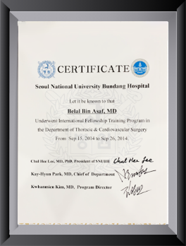
Dr. Belal Bin Asaf's training includes experience as a Clinical Registrar in General Thoracic Surgery at AIIMS, New Delhi, and a thoracic surgery fellowship at Seoul National University Bundang Hospital, South Korea.
For a suspected chest wall tumour, consultation focuses on establishing the diagnosis, reviewing treatment options and determining whether surgery is appropriate. Where resection is needed, planning considers both tumour clearance and the need for reconstruction.
Dr. Belal Bin Asaf holds a monthly visiting outpatient clinic at New City Hospital, Tengpora Bypass, Srinagar.
The visiting clinic provides consultation, examination and review of reports and scans. Further investigations are coordinated according to clinical need. Surgery under Dr. Asaf's care is arranged at Medanta Hospital, Noida.
Please confirm the next OPD date, venue and appointment timings with the appointment team before travelling. An enlarging or concerning lump should be evaluated promptly; patients should not delay assessment solely to wait for the monthly clinic.
Bring previous consultation notes, imaging reports and the actual scan images in digital format where available. If a biopsy has already been performed, bring the pathology report; slides or tissue blocks may be requested for review.
If you have a new or growing chest wall lump, arrange an assessment to understand its cause and the appropriate next step.
Contact the appointment team to book the next Srinagar consultation or arrange an earlier appointment at the Noida centre.
See the full thoracic surgeon in Srinagar OPD details, or book a consultation online.

Medically Written & Reviewed By
Director, Institute of Chest Surgery, Chest Onco-Surgery & Lung Transplantation — trained at AIIMS, New Delhi, with a Thoracic Surgery fellowship at Seoul National University Bundang Hospital, South Korea; first surgeon in India to perform a Robotic Subxiphoid Thymectomy. View full credentials & experience →


Morning: 10:00AM - 2:00PM
Evening 4:00PM - 6:00PM(Mon to Sat)


