

Dr. Belal Bin Asaf
Thoracic (Chest) Surgeon
Director, Institute of Chest Surgery,
Chest Onco-Surgery & Lung Transplantation, Medanta Hospital

-
 [email protected]
-
Follow Us:


Dr. Belal Bin Asaf
Thoracic (Chest) Surgeon
Director, Institute of Chest Surgery,
Chest Onco-Surgery & Lung Transplantation, Medanta Hospital
[email protected]

Patients in Srinagar and elsewhere in Jammu & Kashmir who have a lung nodule, suspected lung cancer, a complex chest infection, recurrent pneumothorax or another condition involving the chest may benefit from an assessment by a thoracic surgeon.
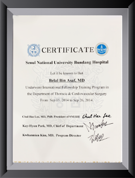
Dr. Belal Bin Asaf is Director of the Institute of Chest Surgery, Chest Onco-Surgery & Lung Transplantation at Medanta Hospital, Noida. He has over 15 years of experience in thoracic surgery, including video-assisted thoracoscopic surgery (VATS) and robotic thoracic procedures. He trained in General Thoracic Surgery at AIIMS, New Delhi, and underwent a thoracic surgery fellowship at Seoul National University Bundang Hospital, South Korea.
He offers scheduled visiting outpatient consultations in Srinagar to review reports and discuss treatment options. Procedures and operations, when needed, are arranged at an appropriate treatment centre; the visiting OPD is not an emergency or surgical service.
Thoracic surgeons assess and operate on selected diseases of the lungs, airways, pleura (the lining around the lungs), mediastinum (the central compartment of the chest), chest wall, diaphragm and oesophagus. Not every patient referred to a thoracic surgeon requires surgery. Many conditions are best managed through joint decision-making with pulmonologists, oncologists, interventional radiologists, gastroenterologists and other specialists.
A thoracic surgical opinion may be useful when a scan identifies a suspicious lung lesion, when infection or an air leak persists despite appropriate treatment, or when a defined anatomical problem could be corrected surgically. The assessment focuses on the diagnosis, available treatment options, operative risks and likely benefits -- not on proceeding to an operation by default.
A lung nodule is a spot or small opacity seen on chest imaging. Many nodules are benign, including scars and healed infections, but some represent early lung cancer. The next step depends on nodule size, appearance, growth over time, smoking and cancer history, and overall clinical risk.
Assessment may involve comparison with earlier CT scans, interval CT surveillance, PET-CT, image-guided biopsy, bronchoscopy or endobronchial ultrasound (EBUS), depending on the case. Not every nodule requires a biopsy or surgical removal. If cancer is diagnosed, staging and multidisciplinary review help determine the safest and most effective approach.
For selected patients with resectable non-small-cell lung cancer, surgery may involve a lobectomy or an appropriately selected anatomical segmentectomy with systematic lymph-node assessment. VATS or robotic surgery may reduce surgical trauma in suitable cases. Other patients benefit from systemic therapy, radiotherapy, perioperative treatment or combinations of these modalities according to stage, biomarkers and fitness.
Coughing up blood can be caused by respiratory infection, bronchiectasis, active or previously treated tuberculosis, aspergilloma, cancer or vascular abnormalities. The cause and severity determine treatment. Initial evaluation commonly involves clinical assessment and chest imaging, often contrast-enhanced CT or CT angiography; bronchoscopy is useful in selected cases.
Many patients are treated medically. Bronchial artery embolisation by an interventional radiologist is an important option for significant or recurrent bleeding. Surgery may be considered when bleeding arises from a localised, resectable area of diseased lung, particularly if episodes persist or recur despite appropriate treatment.
Sudden heavy bleeding, breathing difficulty, fainting or rapidly worsening symptoms require immediate emergency care at the nearest equipped hospital -- not a monthly outpatient appointment.
Bronchiectasis is permanent widening of the airways, which can cause chronic sputum production, recurrent infections and sometimes haemoptysis. It may follow previous tuberculosis, but also has many other causes.
Most patients benefit from specialist medical management, including airway-clearance techniques, treatment of infections and assessment for underlying causes. In carefully selected patients with disease confined to one region of the lung and persistent symptoms despite optimised treatment, surgical resection may be considered. CT imaging, pulmonary function and the extent of disease guide this decision.
Empyema is pus in the pleural space surrounding the lung, usually associated with infection. A complicated parapneumonic effusion is an infected or high-risk fluid collection that may also require drainage. Patients may have fever, pleuritic chest pain, cough and breathlessness.
Treatment usually includes antibiotics and, for established pleural infection, image-guided chest-tube drainage. Thoracic ultrasound, pleural fluid tests and sometimes CT help evaluate the collection. If infected fluid remains despite chest drainage, a combination of intrapleural tPA and DNase may be appropriate in selected patients.
VATS drainage, debridement or decortication may be needed for persistent infection, inadequate drainage or a restrictive pleural peel that prevents lung expansion. Surgery is not automatically necessary for every loculated collection; timing and method should be decided from the full clinical picture.
A pneumothorax occurs when air enters the pleural space and part or all of the lung collapses. It may happen spontaneously, in association with lung disease, or following injury or a medical procedure.
Management is based on symptoms, physiological stability, underlying disease and patient circumstances -- not the size shown on X-ray alone. Selected stable patients can be observed; others may need needle aspiration, ambulatory treatment or chest drainage.
Thoracic surgery may be considered after a second pneumothorax on the same side, a first episode on the opposite side, persistent air leak, bilateral episodes or when recurrence prevention is especially important. VATS bullectomy and pleurodesis are options in suitable patients, but no treatment eliminates recurrence risk entirely.
Sudden breathlessness or severe chest pain requires urgent assessment. Suspected tension pneumothorax is an emergency requiring immediate treatment.
Masses may arise within the mediastinum, thymus gland, ribs, cartilage or chest-wall soft tissues. Some are benign, while others require tissue diagnosis, oncological treatment or resection. The relationship to nearby vessels, nerves and other structures is central to planning.
Selected thymic and mediastinal tumours can be removed using VATS or robotic approaches, whereas large, invasive or complex lesions may require open surgery. Chest-wall tumour surgery may involve removal and reconstruction of part of the chest wall. For suspected thymoma, assessment may also include symptoms or tests for myasthenia gravis.
Biopsy is not always the first step for every mediastinal mass; investigations should be tailored to the suspected diagnosis and performed after appropriate specialist review.
Thoracic surgical services may also address selected oesophageal disorders and tumours, diaphragmatic abnormalities, pleural tumours, airway narrowing and other complex chest conditions. The appropriate investigations and treatment depend on the diagnosis and may involve a multidisciplinary team.
A clear surgical decision requires more than an abnormal CT report. The treating team assesses the diagnosis, disease extent, likely benefit of intervention, previous treatment, medical comorbidities and the patient's preferences.
Depending on the condition, tests may include contrast-enhanced CT, PET-CT, bronchoscopy or EBUS, pulmonary function testing, exercise assessment, cardiac evaluation and tissue diagnosis. For suspected lung cancer, molecular and pathological findings may also influence the overall treatment sequence.
When an operation is appropriate, Dr. Asaf discusses the rationale, minimally invasive versus open approaches, possible complications, recovery expectations and alternatives. VATS and robotic techniques are useful surgical tools -- not substitutes for sound selection and operative planning.
Dr. Asaf's practice includes lung cancer surgery, complex pleural conditions, bronchiectasis and aspergilloma, recurrent pneumothorax, mediastinal tumours and other thoracic surgical diseases. His experience with VATS and robotic thoracic surgery supports assessment of both minimally invasive and open surgical options.
The purpose of the consultation is to provide a practical specialist opinion: whether further tests are needed, whether continued non-surgical care is preferable, and whether an operation could offer meaningful benefit. Coordinated care with the patient's local physician or pulmonologist remains important. Dr. Belal Bin Asaf trained as a Clinical Registrar in General Thoracic Surgery at AIIMS, New Delhi, and completed a fellowship in Thoracic Surgery at Seoul National University Bundang Hospital, South Korea. Read more
Dr. Belal Bin Asaf is scheduled to hold a visiting outpatient consultation at New City Hospital, Tengpora Bypass, Srinagar, on the third Saturday of each month, from 9:00 AM to 11:00 AM and 2:00 PM to 5:00 PM. Please confirm before travel because schedules may change.
The Srinagar visit is for an outpatient examination, second opinion and review of existing scans and treatment records. When further procedures or surgery are required, care may be arranged at Medanta Hospital, Noida, according to medical needs and current institutional arrangements.
Please bring CT or PET-CT images (not only written reports), previous discharge summaries, biopsy or bronchoscopy results, a medication list and relevant lung-function tests. Acute emergencies, including major haemoptysis, severe breathlessness or suspected tension pneumothorax, should be assessed immediately at a nearby emergency hospital.
No. Many nodules require only risk assessment and CT follow-up. Biopsy or resection is considered when imaging and clinical findings indicate a meaningful cancer risk.
Yes, in selected patients with resectable disease. Stage, lymph-node involvement, tumour anatomy, lung function and other treatments determine suitability.
Surgery is considered when appropriate antibiotics and drainage fail, or when organised pleural disease causes persistent infection or restricted lung expansion. Some patients benefit from intrapleural tPA and DNase instead.
Not always. Surgical recurrence prevention is commonly discussed after recurrence and in other higher-risk situations, taking patient preferences and medical fitness into account.
Often, yes. Treatment of infection, bronchoscopy and bronchial artery embolisation can be effective. Surgery is reserved for selected cases.
Yes. The visiting Srinagar OPD is intended for review and planning. If surgery is recommended, the procedure can be arranged at an appropriately equipped centre.
If you have a persistent chest condition, a suspicious lung scan or a recommendation for thoracic surgery, a specialist assessment can clarify the appropriate next step. Arrange an appointment for the visiting Srinagar OPD or at Medanta Hospital, Noida, depending on the urgency and type of evaluation required.
Patient information only. It does not replace personalised medical advice or emergency care. Confirm visiting clinic details before making travel plans.
We are here for you! How can we help?

Thoracic surgeons specialize in surgical procedures involving the chest, addressing a variety of complex conditions related to the heart, lungs, esophagus, and other thoracic organs. Their expertise includes both traditional surgeries and minimally invasive techniques to improve patient outcomes.
Thoracic (Chest) Surgeons specialize in performing surgeries on all the organs in the chest, excluding the heart and major blood vessels. This includes treating diseases affecting the lungs, windpipe (trachea and bronchus), pleura (the thin layer covering the lungs and chest wall), mediastinum (the central part of the chest cavity between the lungs), chest wall, diaphragm, and other related organs. Thoracic Surgeon is skilled in addressing these complex conditions with advanced care.
With over 15 years of experience, Dr. Belal Bin Asaf is a leader in Video-Assisted Thoracoscopic Surgery (VATS), offering patients smaller incisions, less pain, and faster recovery.
Dr. Belal Bin Asaf utilises advanced robotic technology for thoracic surgery, providing enhanced precision and better outcomes through state-of-the-art techniques.
Working with a multidisciplinary team, Dr. Belal Bin Asaf ensures personalised treatment plans and holistic care from diagnosis to recovery.
Dr. Belal Bin Asaf has a history of successful surgeries across a range of thoracic conditions, including lung cancer, thymoma, and chest wall tumors.
Dr. Belal Bin Asaf empowers patients with clear explanations of diagnoses and treatments, fostering informed and confident decision-making.
Choosing Dr. Belal Bin Asaf means opting for expertise, innovation, and compassionate care in thoracic surgery.


Medically Written & Reviewed By
Director, Institute of Chest Surgery, Chest Onco-Surgery & Lung Transplantation — trained at AIIMS, New Delhi, with a Thoracic Surgery fellowship at Seoul National University Bundang Hospital, South Korea; first surgeon in India to perform a Robotic Subxiphoid Thymectomy. View full credentials & experience →


Morning: 10:00AM - 2:00PM
Evening 4:00PM - 6:00PM(Mon to Sat)


