

Dr. Belal Bin Asaf
Thoracic (Chest) Surgeon
Director, Institute of Chest Surgery,
Chest Onco-Surgery & Lung Transplantation, Medanta Hospital

-
 [email protected]
-
Follow Us:


Dr. Belal Bin Asaf
Thoracic (Chest) Surgeon
Director, Institute of Chest Surgery,
Chest Onco-Surgery & Lung Transplantation, Medanta Hospital
[email protected]
Consult Dr. Belal Bin Asaf, Director – Institute of Chest Surgery, Chest Onco-Surgery & Lung Transplantation, Medanta Noida
A chest infection that does not improve despite appropriate antibiotic treatment, particularly when accompanied by persistent fever, chest pain or increasing breathlessness, may indicate a complication known as empyema thoracis.
Empyema is a serious infection in which pus collects in the pleural space surrounding the lung. Unlike an uncomplicated chest infection, established empyema usually requires drainage of the infected collection in addition to antibiotics. Without timely treatment, the infection may become organised, restrict lung expansion and lead to prolonged illness.
Dr. Belal Bin Asaf, an experienced thoracic surgeon, offers specialist consultations for empyema and other complex chest conditions in Srinagar. He serves as Director of the Institute of Chest Surgery, Chest Onco-Surgery & Lung Transplantation at Medanta Hospital, Noida.
With over 15 years of experience in thoracic surgery and extensive expertise in video-assisted thoracoscopic surgery (VATS), Dr. Asaf evaluates patients with complicated pleural infections, recurrent or persistent empyema, and trapped lung.
Patients from Srinagar and across Jammu & Kashmir can seek a specialist thoracic surgical opinion locally. When advanced drainage procedures or surgery are required, further evaluation and treatment can be arranged at Medanta Hospital, Noida.
The lungs are surrounded by two thin membranes called the pleura. The narrow space between these membranes normally contains a small amount of fluid, allowing the lungs to move smoothly during breathing.
In certain infections, particularly pneumonia, fluid may accumulate in this space. This is called a parapneumonic pleural effusion.
Some uncomplicated parapneumonic effusions resolve with antibiotic treatment alone. However, if bacteria invade the pleural fluid, the infection may progress to a complicated parapneumonic effusion or empyema.
Empyema thoracis refers to the presence of pus in the pleural cavity.
As the infection progresses, fibrin deposits and internal partitions may develop, dividing the fluid into multiple pockets. Over time, a thick fibrous membrane may form around the lung, preventing it from expanding normally. This condition is commonly called a trapped lung or entrapped lung, depending on the underlying mechanism.
Established empyema is a potentially serious condition requiring prompt medical assessment, appropriate antibiotics and, in most cases, drainage of the infected pleural collection.
Symptoms vary according to the severity and duration of infection.
Common symptoms include:
Not every patient develops all these symptoms. Older adults and people with diabetes or impaired immunity may have less obvious signs of infection.
A patient who initially improves with antibiotics but subsequently develops recurrent fever, chest discomfort or increasing breathlessness should be evaluated for possible pleural infection.
Severe or rapidly worsening breathlessness, low oxygen levels, confusion, marked weakness, persistent high fever or suspected sepsis require immediate hospital assessment.
Patients with these symptoms should not wait for a scheduled outpatient consultation.
Empyema most commonly occurs as a complication of bacterial pneumonia. However, it can develop even when appropriate treatment has already been started.
Other causes and associated conditions include:
Patients with diabetes, impaired immunity, chronic kidney disease, alcohol dependence or other significant medical illnesses may be more susceptible to severe pleural infections.
However, empyema can also occur in otherwise healthy individuals.
Tuberculosis is an important consideration in patients with persistent or chronic pleural infection, particularly in regions where tuberculosis remains prevalent.
Tuberculous empyema differs from the more common tuberculous pleural effusion. It involves infection of the pleural space with Mycobacterium tuberculosis and may be associated with a thick pleural peel, calcification, persistent infection or bronchopleural fistula.
Treatment requires appropriate anti-tubercular medication. Selected patients may also need drainage or surgery, depending on the extent of the disease and associated complications.
Early diagnosis is important because treatment becomes more complex when infected fluid develops multiple loculations or a thick fibrous covering.
Diagnosis involves clinical examination, chest imaging and, where appropriate, analysis of pleural fluid.
A chest X-ray is often the first investigation performed. It can identify fluid around the lung, associated pneumonia or reduced expansion of the affected lung.
However, an X-ray alone cannot reliably determine whether pleural fluid is infected.
Chest ultrasound is particularly useful in evaluating pleural collections.
It helps assess:
Ultrasound guidance improves the safety and accuracy of pleural procedures.
A contrast-enhanced CT scan may be recommended when the infection is extensive, the anatomy is complex or an underlying cause needs to be identified.
CT imaging can demonstrate pleural thickening, loculated collections, associated lung disease and findings that may influence surgical planning.
When appropriate, a sample of fluid is obtained through ultrasound-guided thoracentesis.
Investigations may include:
Frank pus in the pleural cavity establishes the diagnosis of empyema.
In non-purulent parapneumonic effusions, a pleural fluid pH of 7.2 or below strongly supports the need for drainage when adequate safe access is available.
Other findings, including low glucose, elevated LDH, positive microbiology and complex ultrasound appearances, may also influence the decision.
Not every pleural effusion requires drainage. The need for intervention depends on clinical findings, fluid characteristics and the patient's response to treatment.
Empyema is traditionally described in three stages. Understanding these stages helps explain why some patients respond to drainage and antibiotics while others require surgical treatment.
However, these stages may overlap. Treatment decisions depend on the patient's condition, imaging findings and response to treatment rather than the stage alone.
This is the early phase, when fluid begins accumulating in the pleural cavity.
The fluid is generally thin and free-flowing, with limited internal organisation.
An uncomplicated parapneumonic effusion may respond to antibiotics alone. If the collection is infected or meets criteria for drainage, a chest tube may be necessary.
As the infection progresses, fibrin deposits develop and the pleural fluid may become more viscous.
Internal partitions divide the fluid into multiple pockets, known as loculations, which can make drainage difficult.
Treatment may involve image-guided chest-tube drainage, intrapleural medication or VATS, depending on the extent of infection and response to initial management.
In advanced disease, a thick fibrous membrane, commonly called a pleural peel, may develop over the lung.
This membrane can restrict expansion even after the infected fluid has been drained.
Patients with persistent infection, inadequate drainage or significant restriction of lung expansion may benefit from surgical decortication.
Not every patient in the organising stage requires surgery. The decision depends on symptoms, residual infection, lung expandability and the patient's fitness for an operation.
The main objectives of empyema treatment are to control infection, remove infected pleural fluid and restore lung expansion as effectively as possible.
Treatment is individualised according to the severity of infection, imaging findings, associated medical conditions and response to initial treatment.
Antibiotics are an essential part of managing pleural infection.
Treatment is initially selected according to the likely organisms, whether the infection was acquired in the community or in hospital, and local antibiotic resistance patterns.
Antibiotics may subsequently be adjusted based on microbiological results and clinical response.
The duration of treatment varies and often extends over several weeks, particularly in patients with extensive or complicated infections.
While uncomplicated parapneumonic effusions may resolve with antibiotics alone, established empyema generally requires drainage in addition to antimicrobial treatment.
When infected pleural fluid or pus is present, drainage is usually required.
A chest tube is inserted into the pleural space, often under ultrasound guidance, allowing infected fluid to drain.
Small-bore chest tubes are effective for initial drainage in many patients.
The medical team monitors clinical improvement, drainage output, inflammatory markers and imaging findings to determine whether additional treatment is necessary.
Some patients have pleural collections divided into multiple pockets, making drainage through a chest tube difficult.
In selected cases, a combination of two medicines—tissue plasminogen activator (tPA) and DNase—may be administered through the chest tube.
These medicines help break down fibrinous septations and reduce the viscosity of infected pleural fluid, improving drainage.
Intrapleural tPA and DNase may reduce the need for surgery in appropriately selected patients.
However, this treatment is not suitable for everyone. Potential bleeding risks, contraindications, the extent of disease and the patient's clinical condition must be considered.
Patients with persistent collections or ongoing infection despite appropriate treatment may require thoracic surgical evaluation.
VATS is a minimally invasive surgical approach used to treat selected patients with complicated or persistent empyema.
The procedure is performed under general anaesthesia using a camera and specialised surgical instruments introduced through small incisions in the chest wall.
Depending on the extent of disease, the operation may involve:
VATS may be considered when initial drainage is inadequate, infected collections persist, the patient is not improving satisfactorily or organised pleural disease prevents lung expansion.
Compared with open surgery, VATS may offer reduced surgical trauma, less postoperative discomfort and a shorter hospital stay in appropriately selected patients.
However, the choice of procedure must prioritise safe and complete treatment of the infection rather than the size of the incision alone. With an experienced surgical team majority of Empyema Thoracis surgery i.e. decortication can be done by VATS.
In some patients, particularly those with longstanding organised empyema, dense adhesions or extensive pleural fibrosis, minimally invasive surgery may not provide adequate clearance.
An open thoracotomy may then be required.
During decortication, the surgeon removes the restrictive fibrous pleural peel where necessary to allow the underlying lung to expand.
The decision between VATS and open surgery depends on the extent of disease, anatomical findings, patient condition and the ability to achieve adequate surgical treatment.
In selected patients, an operation initially attempted through VATS may require conversion to an open procedure.
Not every patient with empyema requires surgery.
Many patients improve with appropriate antibiotics, chest-tube drainage and, where indicated, intrapleural therapy.
Thoracic surgical evaluation becomes particularly important when:
Early involvement of a thoracic surgeon can help determine whether continued drainage-based treatment or surgical intervention offers the best chance of recovery.
The goal is not to operate on every patient, but to avoid unnecessary delays when surgery is clinically indicated.
Untreated pleural infection can lead to progressively organised collections, persistent sepsis, prolonged hospitalisation and impaired lung expansion.
As fibrin deposits and pleural thickening develop, simple drainage may become less effective.
Early recognition and appropriate treatment can reduce the risk of progression and help prevent avoidable complications.
However, early treatment does not guarantee that surgery will be unnecessary. Some patients develop complicated infections despite prompt medical care.
The most important principle is timely reassessment and escalation of treatment when the initial approach is not producing satisfactory improvement.
Recovery depends on the severity and duration of infection, the patient's general health and the treatment required.
Patients treated with antibiotics and chest-tube drainage may recover without an operation, although hospitalisation and antibiotic treatment can still be prolonged.
Following VATS or open decortication, a chest drain is generally retained until drainage is satisfactory and the lung has adequately re-expanded.
Postoperative care may include:
VATS can offer faster recovery than open thoracotomy in appropriately selected patients.
However, recovery time varies considerably. Patients with advanced infection, prolonged illness, poor nutritional status or significant underlying lung disease may require a longer period of rehabilitation.
Full lung expansion and improvement in respiratory function depend on the extent of pre-existing damage and the success of infection control.
Empyema management requires careful assessment of the extent of infection, the condition of the underlying lung and the response to initial drainage.
Patients with complex pleural disease may benefit from thoracic surgical expertise, particularly when minimally invasive drainage, debridement or decortication is being considered.
Dr. Belal Bin Asaf is Director of the Institute of Chest Surgery, Chest Onco-Surgery & Lung Transplantation at Medanta Hospital, Noida.
He has over 15 years of experience in thoracic surgery, with extensive experience in video-assisted thoracoscopic and robotic thoracic procedures.
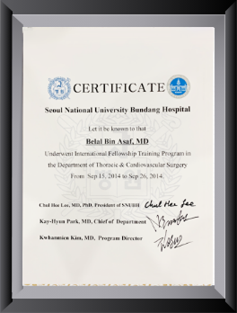
He trained in General Thoracic Surgery at the All India Institute of Medical Sciences (AIIMS), New Delhi, and completed a fellowship in Thoracic Surgery at Seoul National University Bundang Hospital, South Korea.
His thoracic surgical practice includes minimally invasive treatment of complex pleural infections, VATS debridement and decortication, and management of other advanced chest conditions.
Successful empyema treatment often involves collaboration between pulmonologists, infectious disease specialists, radiologists, critical care physicians and thoracic surgeons.
Dr. Asaf's approach focuses on selecting an appropriate treatment for each patient, identifying situations where surgery may be beneficial and avoiding unnecessary delays in managing complicated pleural infection.
Patients seeking an empyema specialist in Srinagar can obtain a thoracic surgical opinion regarding existing investigations, treatment response and the need for further intervention.
Patients from Srinagar and other parts of Jammu & Kashmir can consult Dr. Belal Bin Asaf during his scheduled visiting outpatient clinic.
Consultation timings: 10 AM to 2:00 PM.
Please confirm the latest clinic date, location and timings before travelling, as visiting schedules may change.
The Srinagar consultation is intended for specialist evaluation, review of reports and planning further treatment.
Patients are encouraged to bring their previous chest X-rays, CT scans, ultrasound reports, discharge summaries, pleural fluid investigations and details of antibiotic or chest-tube treatment already received.
When advanced thoracic surgery is required, patients may be referred for treatment at Medanta Hospital, Noida, according to clinical requirements and hospital arrangements.
Important: Patients with severe breathlessness, suspected sepsis, persistent high fever or rapid clinical deterioration should seek immediate treatment at an appropriately equipped local hospital. Urgent drainage and stabilisation should not be delayed while waiting for a visiting specialist consultation.
Yes. Empyema is an infection of the pleural space that may lead to persistent sepsis, impaired lung expansion and other complications if not treated appropriately. Most patients can be treated successfully, but prompt diagnosis and appropriate management are important.
Yes. Many patients recover with antibiotics and appropriate chest-tube drainage. Some may also benefit from intrapleural tPA and DNase. Surgery is considered when these treatments are inadequate or when organised pleural disease requires an operation.
No. Fluid around the lungs is called a pleural effusion and can occur for many reasons, including heart failure, infection, malignancy and other conditions. Empyema specifically refers to pus within the pleural space.
VATS decortication is a minimally invasive thoracic surgical procedure in which a restrictive fibrous membrane is removed from the lung through small incisions using a camera and specialised instruments. It is performed when clinically indicated to help release the lung and improve its ability to expand.
VATS is generally preferred when the infection can be adequately treated through a minimally invasive approach. It may offer advantages in postoperative discomfort and recovery. However, patients with extensive organised disease or dense adhesions may require open surgery.
Recurrence is possible, although successful drainage and treatment of the underlying infection reduce the risk. Persistent cavities, inadequate drainage, an ongoing source of infection or certain underlying conditions can increase the likelihood of recurrence.
Recovery varies according to the severity of infection, the type of surgery and the patient's general health. Some patients resume routine activities relatively quickly after VATS, while others require several weeks or longer, particularly after advanced infection or prolonged hospitalisation.
Yes. Tuberculosis can cause chronic pleural infection and tuberculous empyema. Treatment generally requires anti-tubercular medication, with drainage or surgery in selected patients.
Not necessarily. Initial evaluation, antibiotics and pleural drainage may be provided at appropriately equipped hospitals in Srinagar. Patients who require advanced thoracic surgical procedures may be referred to Medanta Hospital, Noida, after specialist assessment.
A thoracic surgical opinion is particularly valuable when pleural infection persists despite appropriate antibiotics and drainage, when imaging shows significant loculations or pleural thickening, or when the lung fails to re-expand satisfactorily. Early consultation may help avoid delays if an operation becomes necessary.
Persistent pleural infection, recurrent fluid around the lungs or failure of the lung to expand after drainage should not be ignored.
If you or a family member has been diagnosed with empyema, complicated parapneumonic effusion or trapped lung, a specialist thoracic surgical opinion can help clarify the available treatment options.
Consult Dr. Belal Bin Asaf during his scheduled Srinagar OPD at New City Hospital, Tengpora Bypass, or arrange an appointment at Medanta Hospital, Noida, for further evaluation.
The objective is to provide timely, evidence-based treatment, control infection and preserve lung function wherever possible.
See the full thoracic surgeon in Srinagar OPD details, or book a consultation online.
This information is intended for patient education and does not replace an individual clinical assessment. Patients with severe symptoms should seek urgent hospital care.

Medically Written & Reviewed By
Director, Institute of Chest Surgery, Chest Onco-Surgery & Lung Transplantation — trained at AIIMS, New Delhi, with a Thoracic Surgery fellowship at Seoul National University Bundang Hospital, South Korea; first surgeon in India to perform a Robotic Subxiphoid Thymectomy. View full credentials & experience →


Morning: 10:00AM - 2:00PM
Evening 4:00PM - 6:00PM(Mon to Sat)


