

Dr. Belal Bin Asaf
Thoracic (Chest) Surgeon
Director, Institute of Chest Surgery,
Chest Onco-Surgery & Lung Transplantation, Medanta Hospital

-
 [email protected]
-
Follow Us:


Dr. Belal Bin Asaf
Thoracic (Chest) Surgeon
Director, Institute of Chest Surgery,
Chest Onco-Surgery & Lung Transplantation, Medanta Hospital
[email protected]


When pneumonia doesn't fully clear and pus begins collecting around the lung, the result is empyema — one of the more serious conditions a chest surgeon manages, precisely because early recognition and correctly staged treatment make the difference between a short recovery and a much longer, more invasive one. This kind of complex chest infection is treated by Dr. Belal Bin Asaf, Director of the Institute of Chest Surgery, Chest Onco-Surgery & Lung Transplantation at Medanta Hospital, Noida (Sector 50), who brings more than 15 years of thoracic surgery experience and specializes in robotic thoracic surgery, VATS, lung cancer, and myasthenia gravis & thymoma treatment.
Here's a full picture of the condition, how it progresses, and how it's diagnosed and treated — including where nearby patients, and the empyema specialist Gurgaon families turn to, can be reached without unnecessary delay.
Every lung sits within a thin, two-layered pleural lining, and the narrow space between those layers is meant to stay essentially empty aside from a small amount of lubricating fluid. Empyema develops when that space instead fills with infected fluid — thick, pus-laden, and inflammatory — most commonly as a direct extension of pneumonia. The lung infection spreads outward into the adjoining pleural space, converting what might otherwise be a simple parapneumonic effusion into something far harder to treat. Once this happens, the problem is no longer confined to the lung tissue itself; the fluid and the debris it carries begin to interfere mechanically with lung function.
Clinically, empyema is understood in three sequential stages. It starts as the exudative stage — thin, infected fluid that is generally still manageable with drainage alone. Left to progress, it becomes fibrinopurulent, as strands of fibrin build up within the fluid and partition it into distinct, harder-to-access loculated pockets. The final stage is the organizing stage, in which a thick fibrous peel forms across the lung surface, physically trapping the lung and preventing it from fully re-expanding even after the active infection subsides. The most frequent cause is pneumonia that was treated too late, or with an antibiotic course inadequate to the organism involved; other contributing factors include post-operative chest infections, trauma, and infection spreading from elsewhere in the body.
The presentation often mimics stubborn pneumonia: persistent or recurring fever with chills, chest pain that's frequently worse on deep breathing or coughing, breathlessness, an ongoing cough, and pronounced fatigue. Perhaps the most telling clue is timing — a patient already on antibiotics for pneumonia who isn't improving as expected after several days. That lack of response is frequently what leads a physician to investigate specifically for an evolving empyema rather than simply extending the antibiotic course.
Assessment usually begins with a chest X-ray to identify fluid collecting around the lung, followed by ultrasound or CT imaging of the chest to characterize that fluid in detail — critically, to determine whether it remains free-flowing or has already become loculated into separate pockets. Thoracentesis, in which fluid is drawn out with a needle for laboratory analysis, is the step that confirms the diagnosis and stage: pH, glucose, and LDH levels together with gram stain and culture results tell the surgical team exactly what they're dealing with and how urgently intervention is needed.
Management is tailored to how far the disease has progressed. Simple, early-stage (exudative) empyema frequently resolves with intravenous antibiotics combined with chest tube drainage. Once the disease has advanced to the fibrinopurulent stage, with multiple loculated pockets, or to the organizing stage, with a thick restrictive peel, a drainage tube alone is typically no longer sufficient — surgical removal of the infected material becomes necessary. This is the core of Dr. Belal Bin Asaf's surgical expertise: VATS (video-assisted thoracoscopic surgery) decortication, a minimally invasive, camera-guided technique that clears the pus and the fibrous peel and allows the lung to fully re-expand, generally preferred over open thoracotomy for its smaller incisions and faster recovery whenever the case allows it. Delaying surgery when it's genuinely needed risks the infection becoming chronic and the lung sustaining permanent functional damage.
For patients across Gurugram searching for empyema treatment in Gurgaon, this level of surgical expertise is available locally, right at Medanta – The Medicity, Sector 38. As Director of the Institute of Chest Surgery, Chest Onco-Surgery & Lung Transplantation, Dr. Belal Bin Asaf brings more than 15 years of dedicated thoracic surgical experience, with a particular focus on robotic and minimally invasive procedures — including being the first surgeon in India to perform a robotic subxiphoid thymectomy. That level of technical specialization is directly relevant to empyema care, where correctly judging when drainage is enough versus when VATS decortication is required, and executing that surgery precisely, has a real bearing on how completely the lung recovers. For patients searching for an empyema doctor in Gurgaon, or the best pulmonologist for empyema Gurgaon has to offer, that surgical precision is exactly why they are typically referred straight to Dr. Belal Bin Asaf for the operative stages of care.
Medanta – The Medicity in Sector 38 sits centrally for patients across Gurugram, including those in the DLF phases, Golf Course Road, Sohna Road, and Manesar, as well as residents of South Delhi who often find Medanta more accessible than hospitals on the far side of Delhi. Because empyema care often involves urgent imaging, same-visit thoracentesis, and, when needed, a swift transition to surgery, having every stage of diagnosis and treatment available on a single campus in Sector 38 removes delays that can otherwise cost valuable time in a fast-moving infection. It's this combination that makes Medanta – The Medicity a genuine empyema hospital in Gurgaon, not just a referral point.
If pneumonia isn't improving as expected, or a scan has already shown fluid buildup around the lung, don't leave it unaddressed. Contact Medanta – The Medicity, Sector 38, Gurugram, to arrange a consultation with Dr. Belal Bin Asaf — the empyema specialist Gurgaon patients trust — and get a clear plan for treatment before the condition has a chance to progress.
Prefer to book a consultation online right away? Dr. Belal Bin Asaf also sees patients for this condition in Noida and Delhi. Learn more about his full practice as thoracic surgeon in Gurgaon.


Medically Written & Reviewed By
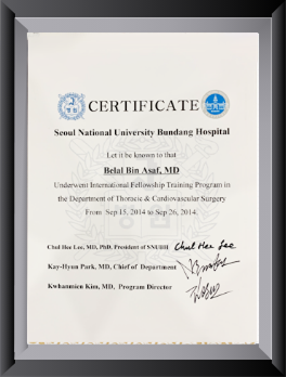
Director, Institute of Chest Surgery, Chest Onco-Surgery & Lung Transplantation — trained at AIIMS, New Delhi, with a Thoracic Surgery fellowship at Seoul National University Bundang Hospital, South Korea; first surgeon in India to perform a Robotic Subxiphoid Thymectomy. View full credentials & experience →


Morning: 10:00AM - 2:00PM
Evening 4:00PM - 6:00PM(Mon to Sat)


