

Dr. Belal Bin Asaf
Thoracic (Chest) Surgeon
Director, Institute of Chest Surgery,
Chest Onco-Surgery & Lung Transplantation, Medanta Hospital

-
 [email protected]
-
Follow Us:


Dr. Belal Bin Asaf
Thoracic (Chest) Surgeon
Director, Institute of Chest Surgery,
Chest Onco-Surgery & Lung Transplantation, Medanta Hospital
[email protected]


When pneumonia doesn't fully clear and pus begins collecting around the lung, the result is a condition called empyema — a serious but treatable problem that needs prompt, expert care. For residents of Noida and the surrounding NCR belt, empyema treatment in Noida is available at Medanta Hospital, Sector 50, under the direction of Dr. Belal Bin Asaf, Director of the Institute of Chest Surgery, Chest Onco-Surgery & Lung Transplantation at Medanta Hospital, Noida. Trained as a Clinical Registrar in General Thoracic Surgery at AIIMS, New Delhi, he brings more than 15 years of focused thoracic surgery experience — spanning robotic thoracic surgery, VATS, lung cancer, and myasthenia gravis & thymoma treatment — to exactly this kind of complex chest infection.
If you or a family member has been battling pneumonia that simply won't respond to antibiotics, or has developed persistent chest pain and fever, this page explains what empyema is, how it progresses, and how it's managed close to home by the empyema specialist Noida families can turn to without traveling outside the NCR.
The pleural space is the thin, normally fluid-free gap between the outer surface of the lung and the inside of the chest wall. Empyema occurs when this space fills with infected fluid — essentially pus — instead of remaining clear. It rarely appears out of nowhere; in the overwhelming majority of cases, it's a downstream complication of pneumonia. Bacteria from an infected lung seed the adjacent pleural fluid, turning what should be a simple, sterile effusion into a thick, infected one. Left unaddressed, this pus doesn't just sit passively — it can scar down and physically restrict how well the lung expands.
Doctors describe empyema as moving through three overlapping stages. It begins as an exudative stage, where the fluid is thin and infected but still relatively easy to drain. If untreated, it advances to a fibrinopurulent stage, during which strands of fibrin crisscross the pleural space, walling the fluid off into pockets, or loculations, that are much harder to reach with a simple tube. Finally, in the organizing stage, a thick fibrous rind forms over the lung's surface, physically trapping it and preventing full re-expansion even after the infection itself is controlled. Beyond inadequately treated pneumonia, empyema can also follow chest trauma, complications after thoracic surgery, or spread from an infection elsewhere in the body.
Most patients present with a pattern that looks like pneumonia refusing to improve: ongoing fever and chills, chest pain that often worsens with breathing or coughing, breathlessness, a persistent cough, and a general sense of fatigue and being unwell. A key warning sign is a patient who has been on appropriate antibiotics for pneumonia for several days but isn't getting better — that plateau, or worsening, is often what prompts a doctor to look specifically for a developing empyema.
Evaluation typically starts with a chest X-ray, which can reveal fluid collecting around the lung. Ultrasound or CT imaging of the chest follows, giving a much clearer picture of whether the fluid is simple or has already broken into loculated pockets. The definitive step is thoracentesis — inserting a needle to draw a sample of the pleural fluid for analysis. Lab testing of this fluid (pH, glucose, LDH levels, and gram stain/culture) tells the treating team not just that infection is present, but how advanced it is, which directly shapes the treatment plan.
Treatment is staged to match the disease. An early, simple (exudative) empyema often responds well to a combination of targeted intravenous antibiotics and chest tube drainage to remove the infected fluid. Once the disease has progressed into the fibrinopurulent or organizing stages, however, a tube alone usually cannot clear the loculated pockets or the fibrous peel encasing the lung — surgery becomes necessary. This is where Dr. Belal Bin Asaf's specific expertise comes in: VATS (video-assisted thoracoscopic surgery) decortication. Using small keyhole incisions and a camera, the infected material and the restrictive peel are meticulously removed and the trapped lung is freed to re-expand, without the larger incision and longer recovery associated with open thoracotomy. Delaying this step carries real risk — chronic pleural infection and permanent loss of lung function are the consequences of empyema left to progress unchecked.
Dr. Belal Bin Asaf is Director of the Institute of Chest Surgery, Chest Onco-Surgery & Lung Transplantation at Medanta Hospital, Noida, with over 15 years of focused experience in thoracic surgery, including advanced minimally invasive and robotic techniques. He was the first surgeon in India to perform a robotic subxiphoid thymectomy, reflecting a broader command of complex, technically demanding chest procedures — the same precision that VATS decortication for empyema demands. For a condition where the difference between early drainage and major open surgery often comes down to how quickly and accurately it's diagnosed and staged, that depth of expertise matters. Many patients begin their search looking for the best pulmonologist for empyema Noida has to offer, only to learn that the surgical stages of this disease are best managed by a thoracic surgeon rather than a pulmonologist alone — which is why he is often the empyema doctor in Noida that referring physicians recommend for advanced, loculated, or organizing-stage disease.
Medanta Hospital in Sector 50 is purpose-built for this level of care and is easily reachable from across the wider Noida-Ghaziabad corridor. Patients from Noida's residential sectors, Greater Noida, and Ghaziabad typically reach the hospital via the Noida Expressway or the connecting sector roads without needing to cross into another city, which matters when a patient is unwell and time-sensitive imaging or a thoracentesis needs to happen quickly. The hospital's full diagnostic and surgical infrastructure — X-ray, CT, ultrasound-guided procedures, and thoracic operating theatres — is on site, so patients don't need to be routed elsewhere between diagnosis and treatment. This combination of round-the-clock diagnostics and on-site thoracic surgery is what makes Medanta Hospital, Sector 50, the region's leading empyema hospital in Noida.
If pneumonia in your family isn't improving the way it should, or a chest X-ray has already flagged fluid around the lung, don't wait for the condition to advance. Contact Medanta Hospital, Sector 50, Noida, to schedule a consultation with Dr. Belal Bin Asaf and get a clear, expert assessment of the right treatment path.
Prefer to book a consultation online right away? Dr. Belal Bin Asaf also sees patients for this condition in Delhi and Gurgaon. Learn more about his full practice as thoracic surgeon in Noida.


Medically Written & Reviewed By
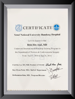
Director, Institute of Chest Surgery, Chest Onco-Surgery & Lung Transplantation — trained at AIIMS, New Delhi, with a Thoracic Surgery fellowship at Seoul National University Bundang Hospital, South Korea; first surgeon in India to perform a Robotic Subxiphoid Thymectomy. View full credentials & experience →


Morning: 10:00AM - 2:00PM
Evening 4:00PM - 6:00PM(Mon to Sat)


