/Adsense//

Introduction: Understanding Lung Nodules

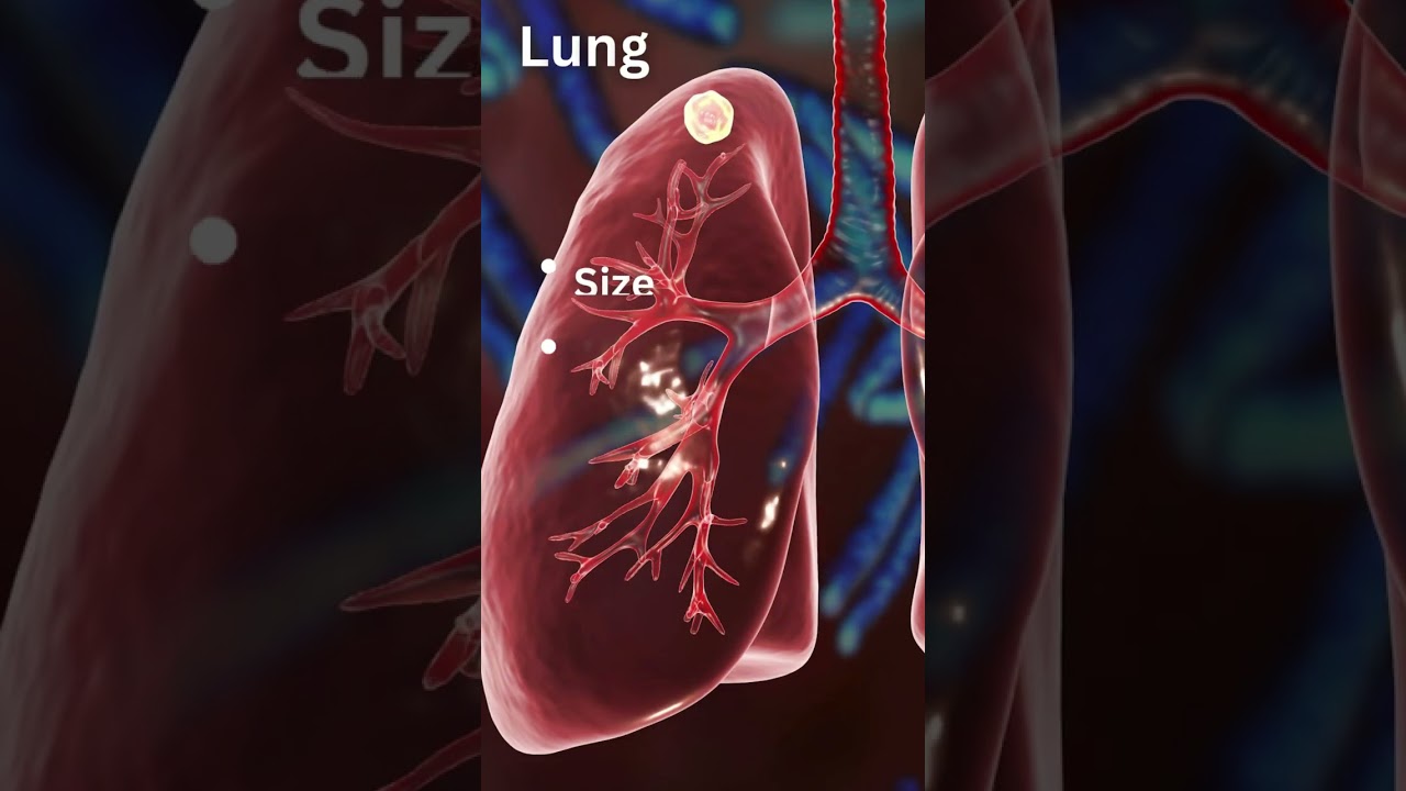
A lung nodule (also called a pulmonary nodule or spot on the lung) is a small, round or oval growth in the lung, usually less than 3 cm in diameter.

Most lung nodules are found incidentally on a chest CT scan done for another reason—such as after a cough, chest pain, or routine health screening.

While the majority of lung nodules are benign (non-cancerous), a proportion can be malignant (cancerous).

Early detection and correct evaluation are crucial for timely and effective treatment.

Key point: Discovering a lung nodule is not automatically a sign of cancer, but it should never be ignored.

Why Lung Nodules Matter

  • Prevalence: Detected in about 1 in 4 chest CT scans.
  • Benign vs Malignant: About 60-70% are benign; 30-40% may need further work-up.
  • Most solitary lung nodules are benign, but a significant proportion—ranging from about 10% to 85%—are malignant, with the exact proportion depending on patient’s age, nodule size, and clinical context (as in under what circumstances was the nodule detected).
  • Cancer Risk Factors:
    • Age > 40
    • Current or former smoker
    • History of cancer elsewhere
    • Larger nodules (> 8 mm), irregular (spiculated) edges
    • Upper-lobe location, faster growth on serial scans

Early evaluation helps identify nodules that require only observation versus those needing biopsy or surgical removal..

Common Causes of Lung Nodules

Lung nodules may arise from a variety of conditions:

  • Infections:
    • Tuberculosis (TB) – common in India and other endemic areas
    • Fungal infections (e.g., Histoplasma, Aspergillus)
    • Post-pneumonia scars
  • Inflammatory/Autoimmune Diseases:
    • Rheumatoid nodules, sarcoidosis, granulomatosis with polyangiitis
  • Benign Tumors:
    • Hamartomas (often contain fat or calcium)
  • Malignant Tumors:
    • Primary lung cancer
    • Metastases (spread from other organs like breast, colon, kidney)
  • Others:
    • Healed granulomas, scar tissue after infection or trauma

Symptoms of Lung Nodules

Most nodules are silent and found incidentally.

However, some patients—especially with malignant nodules—may notice:

  • Persistent cough
  • Coughing up blood (hemoptysis)
  • Shortness of breath
  • Unexplained weight loss or fatigue
  • Chest pain

Remember: Symptoms usually appear when nodules are larger or linked to cancer.

Evaluation & Diagnosis

    CT Scan: First Step

    • High-Resolution CT (HRCT) provides detailed size, shape, margin, density.
    • Fleischner Society Guidelines help decide whether to monitor or investigate further based on:
      • Nodule size
      • Growth over time
      • Patient’s risk profile (smoker vs non-smoker, age, etc.)

    PET-CT

    • Identifies metabolic activity; “hot” nodules are more likely to be malignant.
    • Helps in staging if cancer is suspected.

    Biopsy Methods

    • CT-Guided Needle Biopsy: Usually for nodules near the outer lung.
    • Bronchoscopic Biopsy (including Navigational & Robotic Bronchoscopy): For centrally located nodules.
    • Video-Assisted Thoracoscopic Surgery (VATS) / Robotic Biopsy:
      • Offers precise and minimally invasive sampling or complete removal.
      • Particularly useful when needle/bronchoscopic biopsy is inconclusive or not feasible.

Observation vs Intervention

    When Observation is Safe

    • Low-risk patients
    • Nodules < 6 mm or calcified
    • Stable size over 2 years

    Surveillance Protocol:

    • Interval CT scans (usually at 3-6, 12, and 24 months).
    • Monitoring for any change in size, shape, or density

    When to Intervene

    • Nodule > 8 mm or with suspicious features
    • Growth on serial scans
    • Patient at high risk for cancer
    • Symptoms present (e.g., hemoptysis)

    Intervention may be in the form of biopsy or surgical removal.

Treatment Options

    For Benign Nodules

    • Infections: Treated with antibiotics or anti-TB / anti-fungal drugs
    • Inflammatory Nodules: Managed with appropriate therapy depending on cause.
    • Stable Benign Nodules: Usually require observation only

    For Malignant or Suspicious Nodules

    • Minimally Invasive Surgery:
      • VATS or Robotic-Assisted Wedge Resection / Segmentectomy
      • Preserves healthy lung tissue while ensuring diagnosis and cure
    • Lobectomy: If cancer is confirmed and localized
    • Multidisciplinary Care: Collaboration between thoracic surgeon, pulmonologist, radiologist, pathologist, and oncologist for optimal outcomes

    Dr Belal Bin Asaf is among India’s leading robotic thoracic surgeons with extensive experience in minimally invasive lung biopsy and cancer surgery.

Prognosis & Follow-Up

    • Most benign nodules remain stable or resolve.
    • Cancer detected early and treated surgically often carries an excellent prognosis.
    • Even after a benign biopsy, periodic follow-up CT scans are essential to ensure no new or recurrent nodules develop.

Addressing Patient Concerns

    • Radiation Exposure: Modern low-dose CT minimizes risk.
    • Biopsy Safety: Minimally invasive techniques are generally safe; risks like bleeding or air leak are rare and manageable.
    • Surgery Recovery: Most patients after VATS or robotic procedures go home in 2-3 days and recover quickly.
    • Cancer Fear: Most nodules are not cancer; timely evaluation brings peace of mind.

FAQs – People Also Ask

No. Most lung nodules are benign. Careful evaluation distinguishes harmless nodules from cancerous ones.

Nodules > 8 mm or those that grow or have irregular edges warrant further testing.

Cancerous nodules often grow noticeably within 1–6 months. Benign nodules typically remain stable.

No. CT helps identify suspicious nodules, but biopsy is needed for a definitive diagnosis.

Modern biopsy techniques are minimally invasive and usually performed under local or general anesthesia with low risk.

Follow-up depends on size, appearance, and risk profile—typically every 3–12 monthsas per guidelines.

Most patients recover in a few days, with minimal pain and quicker return to normal activity compared to open surgery.

Risk is lower—especially for small nodules—but cancer is still possible; evaluation is essential.

Most patients spend 5–7 days in hospital after surgery. Recovery is faster with minimally invasive techniques like VATS or robotic surgery. Long-term, patients experience significant improvement in symptoms and resolution of hemoptysis.

If the aspergilloma is completely removed, recurrence is very rare. However, patients with underlying lung disease remain at risk for other complications and need regular follow-up.

If untreated, aspergilloma can lead to:
  • Recurrent or massive life-threatening hemoptysis
  • Chronic lung infections
  • Severe decline in quality of life

Aspergilloma surgery can be technically challenging because of dense adhesions and fragile blood vessels. However, in experienced thoracic surgery centers, surgical success rates are high with excellent long-term outcomes.

Why Choose Dr Belal Bin Asaf

    • Extensive Experience: 15+ years in thoracic surgery
    • Pioneer in Robotic Thoracic Surgery in India
    • Expertise inminimally invasive diagnosis and removal of lung nodules
    • Leader inmultidisciplinary lung cancer management
    • Compassionate, patient-centric care with focus on safety and rapid recovery

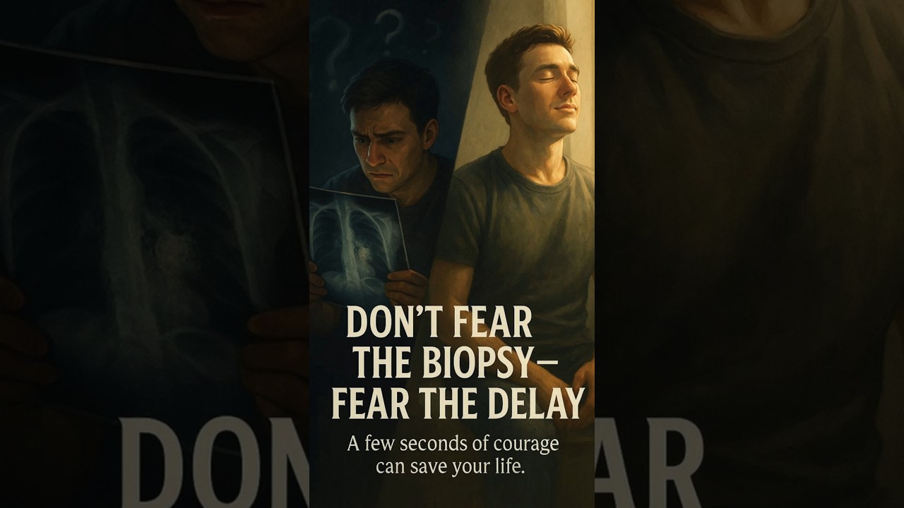
If you or a loved one has been diagnosed with a lung nodule, do not delay evaluation. Early consultation with a thoracic surgeon can make the difference between simple observation and life-saving intervention.

Book a Consultation with Dr Belal Bin Asaf at Medanta

(In-person and virtual appointments available)

References

    • Fleischner Society Pulmonary Nodule Guidelines
    • NCCN Guidelines for Lung Cancer Screening & Diagnosis
    • ACCP Evidence-Based Clinical Practice Guidelines